IOCT

Enhance your possibilities

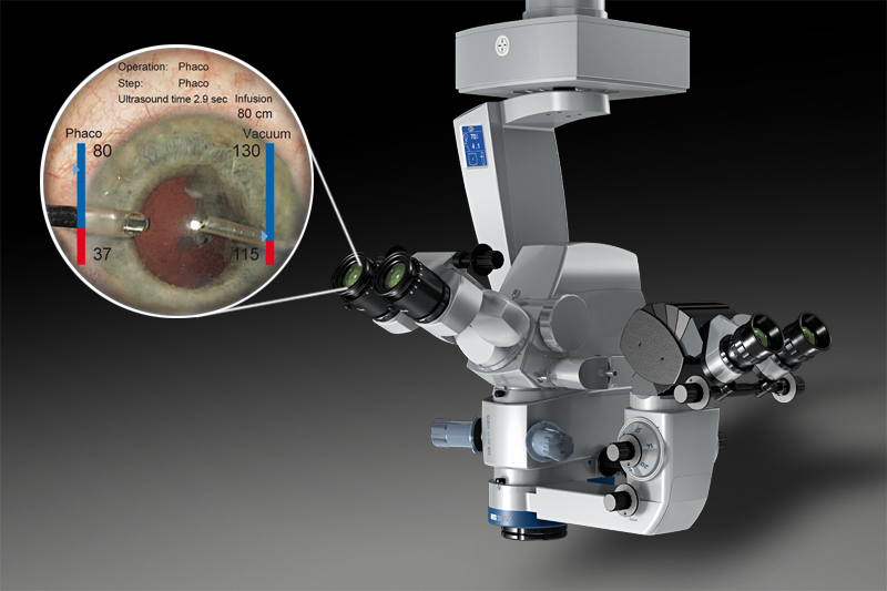
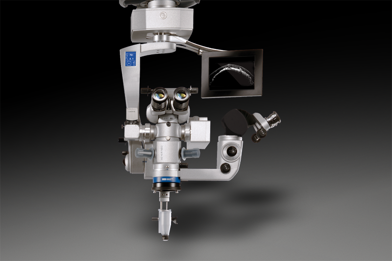
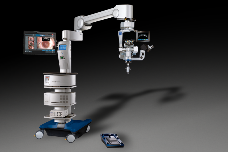
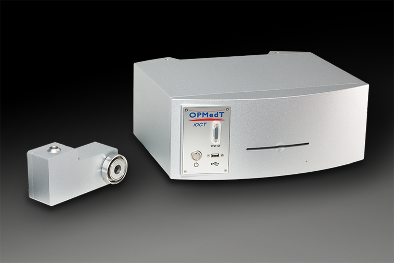
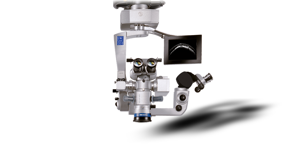
The iOCT camera is integrated into the high class operating microscope HS Hi-R NEO 900A NIR and its carrying system. iOCT scans of the eye’s anterior and posterior segments are performed live during surgery, thus improving quality control mechanisms for the surgeon. Discover more details than with a standard surgical microscope and enhance your possibilities.
APPLICATION
APPLICATION

Experiences with intraoperative OCT

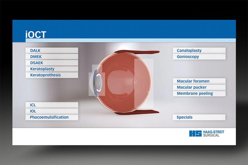
To summarize many iOCT applications HAAG-STREIT SURGICAL has proudly developed an Internet web application. View videos, 3D scans, and picture-in-picture superimpositions from the iOCT-Atlas. Please register free of charge and participate in HAAG-STREIT SURGICAL‘s new world of iOCT.

ANTERIOR SEGMENT SURGERY

Great orientation and detail

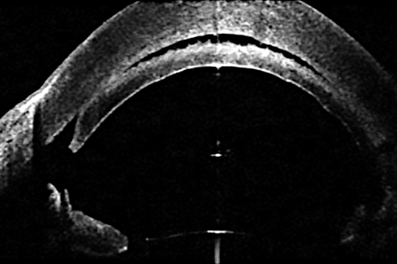
Intraoperative OCT can be used for all surgeries performed in the eye’s anterior segment. While in cataract surgery it is used to help position the IOL, it comprehensively covers all aspects of corneal surgery.

ANTERIOR SEGMENT SURGERY
POSTERIOR SEGMENT SURGERY

Great membrane resolution

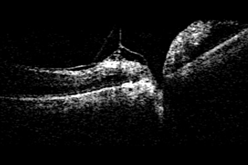
For posterior segment surgery and observation of the retina, intraoperative OCT can be used with HAAG-STREIT SURGICAL’s ophthalmoscopic system EIBOS 2 NIR or a contact lens.
VISUALIZATION
VISUALIZATION

Instant information

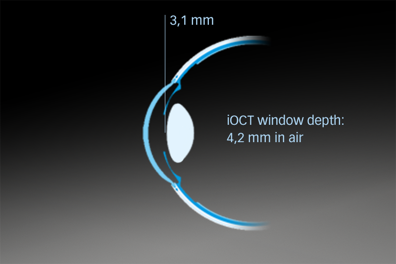
Perfect visualization is supported by the binocular image injection superimposing the scan to the live view into both oculars. The amazing window depth of 4.2 mm provides an overview of the whole anterior segment: from the cornea to the lens.

C.INJECT 900

Enrich your vision

Featuring high definition, the live iOCT image can be superimposed in exceptional resolution into the oculars as an overlay to the live view our image injection system C.INJECT 900. The binocular image injection guarantees distraction-free work in full compliance with your natural, visual perception. Moreover, it prevents drawing your attention away from the surgical field.

C.INJECT 900
HS HI-R NEO 900A
HS HI-R NEO 900A

Ready for iOCT

In order for iOCT observation during surgery to be possible, the microscope needs special optics that are optimized for near infrared (NIR) light. These optics are integrated into the HS Hi-R NEO 900A NIR as well as into the EIBOS 2 NIR. For added flexibility the iOCT-ready microscope system is also offered separately from the actual iOCT, making it possible to upgrade later on.

HANDLING
HANDLING

Intuitive control

For easy handling the intraoperative OCT was designed to focus on the same focal plane as the microscope. In addition, the microscope’s zoom can be used to magnify the iOCT’s image. Further handling is smoothly integrated into the operating microscope, via M.DIS or foot switch.

M.DIS

Microscope mounted display and control
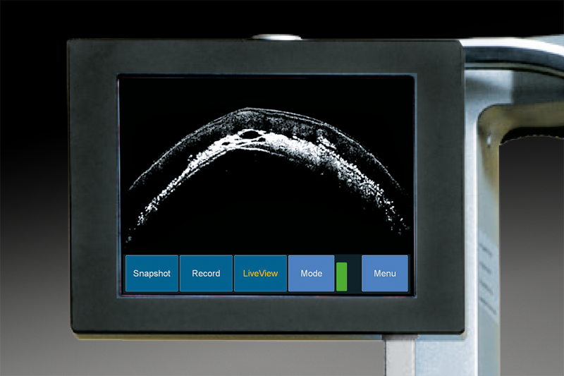
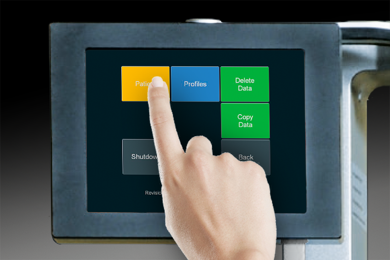
The M.DIS (Microscope Display) operates as the iOCT’s control center. The high-resolution screen, mounted close to the eyepiece, provides the iOCT images which may be seen by momentarily looking up from the eyepiece. The touch screen allows full control of the iOCT including its user settings.
M.DIS
FOOT SWITCH CONTROL
FOOT SWITCH CONTROL

For faster, easier, and safer operation

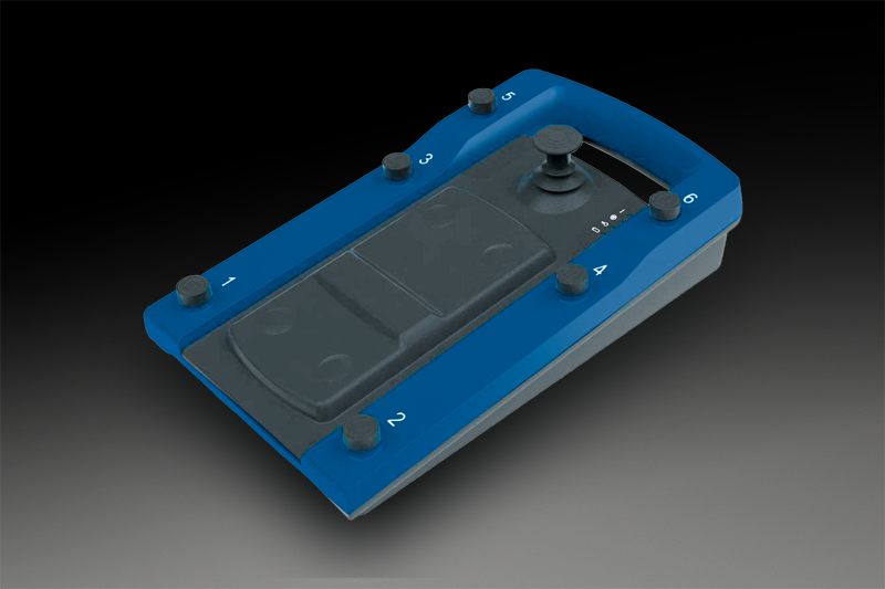
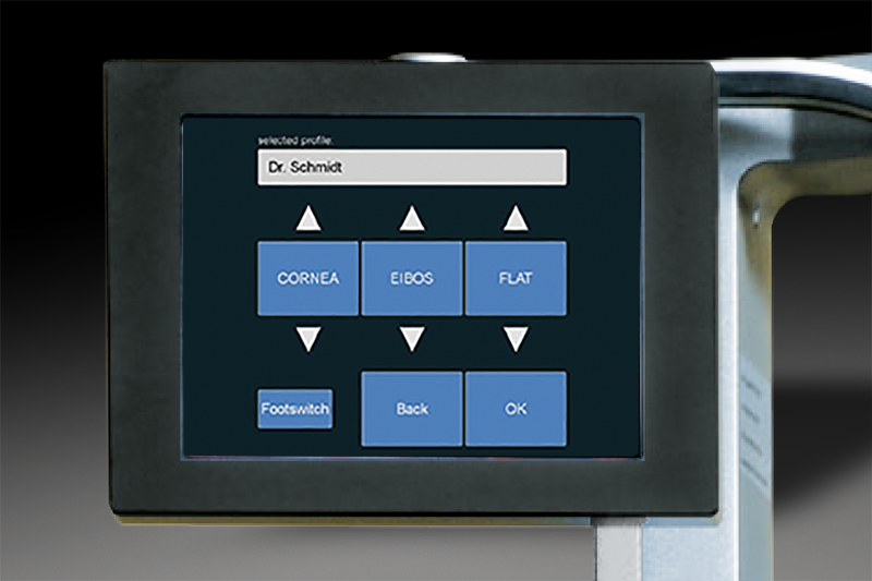
iOCT, together with the operating microscope HS Hi-R NEO 900A NIR, features ergonomic integration of iOCT control into your foot-switch-centered workflow, greatly improving speed and ease of operation. To meet the user experience of established HAAG-STREIT SURGICAL microscopes, the actual assignment of iOCT functionality to foot switch controls is highly customizable through user profiles

USER INTERFACES

Individual workflow optimization

The concept of “per-user customization” is carried far beyond simple footswitch-control mappings. Comprehensive sets of iOCT configurations can seamlessly be saved. These include foot switch operation but also control of special settings related to the eye’s segment observed with iOCT.
USER INTERFACES
RECORDING
RECORDING

Documentation made easy

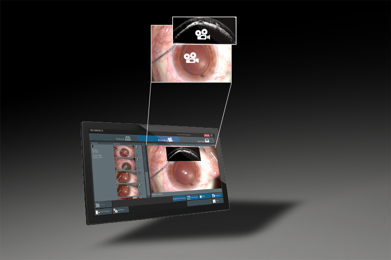
Next to your normal HD camera recording iOCT videos, snapshots, and 3D volume scans need to be made for documentation purposes. This process is fastened by HS MIOS 5 and the recording option M.REC 2 allowing synchronizing these two parallel video signals.

M.REC 2

Connect your recordings

By activating the M.REC 2 the live streams two camera inputs are shown simultaneously on the HS MIOS 5: one as a full screen image, the other displayed as a picture-in-picture. Starting recording or doing a snapshot will automatically activate the function for both streams. This will result in two separate full HD videos being recorded or two separate high quality snapshots being taken.

M.REC 2
VOLUME SCAN
VOLUME SCAN

3-dimensional images

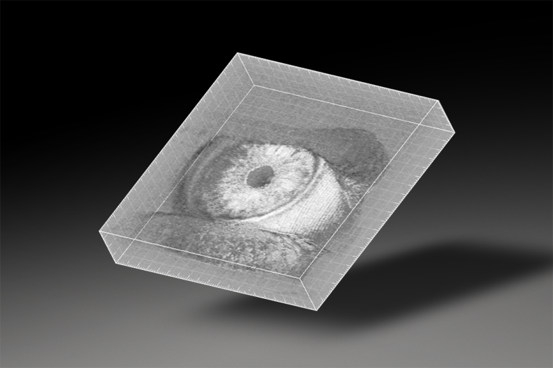
Additionally to your standard recordings, volume scans can be performed with the iOCT system. This offers the unique option of creating a 3-dimensional image including the iOCT‘s depth information during surgery. It is created out of 30 scans over a specified area and clearly supports understanding.
HS MIOS 5

Comprehensive yet intuitive recording
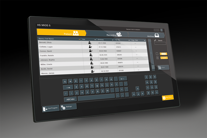
The prime functions of HS MIOS 5 are the recording of operation scenes as well as the capturing and recording of snapshots, together with proper identification of patient and hospital data. Images and video streams can be stored on DVD-R/-RW, HDD, USB flash drive, and external USB hard disk drive or transmitted to the hospital PACS via DICOM.
HS MIOS 5
C.MOR HD / HD³
C.MOR HD / HD³

Compact HD camera

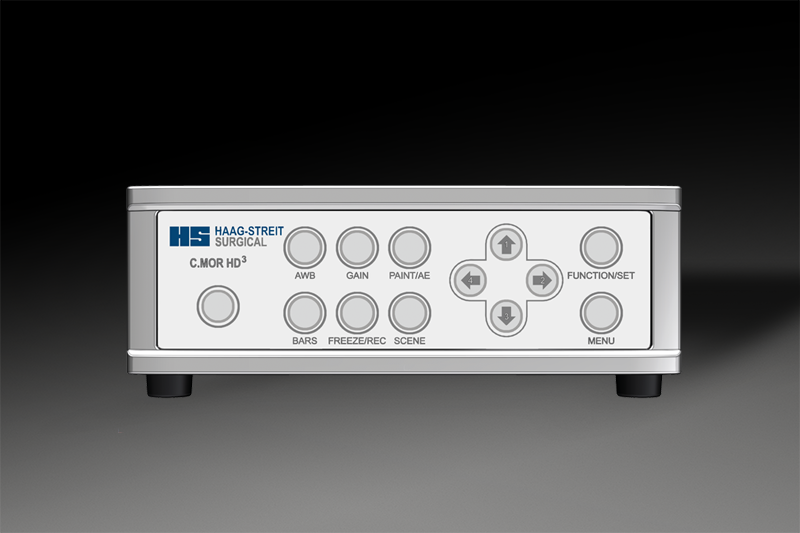
C.MOR HD is a full HD color video camera designed for the HAAG-STREIT SURGICAL microscope. Its ultra-compact camera head houses a 1/3” CMOS sensor that meets very high standards. In its 1 and 3-chip version, images are crisp and sharp. Different user settings allow individual adjustments.
THE PRODUCT’S DEVELOPMENT

The road to success

Already in 2010 the first intraoperative OCT system was demonstrated. Today the iOCT is an established product by HAAG-STREIT SURGICAL that constantly gains importance. When used for teaching, the iOCT images will help trainees learn faster as another dimension is added to their vision.

OPMEDT

The story of iOCT begins

The iOCT was designed by OPMedT GmbH – a member of the HAAG-STREIT family of companies and highly qualified research group in Lübeck, Germany. Capable of integration into optical imaging equipment, a prototype was demonstrated at WOC 2010. Only one year later the product was CE certified. In the following years further technology was integrated. In 2015 510(k) clearance was realized.

OPMEDT