CENTRAL FIELD STATIC PERIMETRY

Covering your essential needs

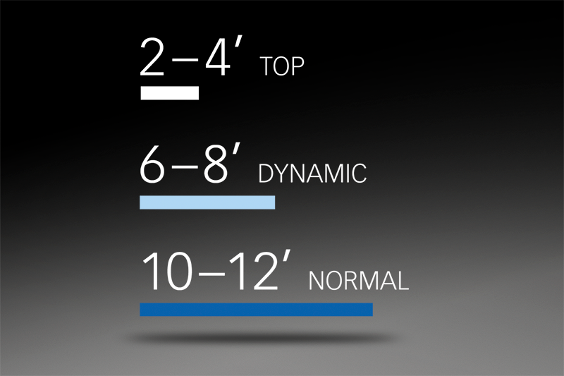
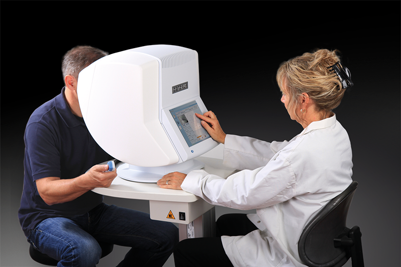
The Octopus 600 performs standard white-on-white threshold testing in just 2–4 minutes in the central visual field. With its comprehensive test library for central tests including G, 32, 30-2, 24-2, M, and 10-2 and its flexible printouts both in Octopus and HFA-format it covers your essential clinical needs. 

STANDARD TEST LIBRARY

The central tests you need

The Octopus 600 offers the most commonly used static tests. For central field testing there are the physiology-based G-patterns following the retinal nerve fibers and the 32, 30-2 and 24-2 patterns. For the macula, there are the physiology-based M pattern and the 10-2 pattern. With the fast TOP strategy, full threshold testing can be completed in just 2-4 minutes.
STANDARD TEST LIBRARY

Tendency-Oriented Perimetry (TOP) presents a further optimisation in fast-threshold testing by reducing the examination time by nearly 80% to just 2–4 minutes compared to 6–8 minutes (dynamic strategy) or 10–12 minutes (normal strategy). The TOP algorithm is a systematic method which takes into account the correlation of the threshold values in neighbouring locations. Since the first test points are presented at a supra-threshold level, even inexperienced patients quickly understand the nature of the test.

All Octopus perimeters offer two unique physiology-based patterns: the G-Program (a 30-degree field for glaucoma assessment) and the M-Program (a 10-degree field for analysing the macula). They are both correlated with a nerve fibre bundle map and thus make it possible to test the points which are most important for a structure-function correlation. These examination patterns offer a higher density of stimuli in the centre, which aids the discovery of paracentral scotomas that are often missed by the common 32 pattern.

The Octopus 600 provides you with the essential stimuli you need. Besides standard white-on-white perimetry with a Goldmann III stimulus, it also offers Pulsar perimetry for early glaucoma detection and corresponding normative databases for each of those stimuli.
OCTOPUS PRINTOUT

Standard Octopus representations

Look at visual field results the way you are used to from your Octopus perimeter. All Octopus perimeters offer the standard 7-in-1 printout with its well-known representations, a customizable 4-in-1 printout, a serial printout and much more. And why not conveniently view results in your office by networking your Octopus to the EyeSuite software on your computer?
OCTOPUS PRINTOUT
HFA-STYLE PRINTOUT
HFA-STYLE PRINTOUT

Smooth transition from HFA

Enjoy a smooth transition from a HFA to an Octopus perimeter. All Octopus perimeters allow you to import your historic HFA data. Because raw data is imported, you can display your historic and new data in the same format of your choice, either as an HFA-style printout or in the Octopus format. To learn more about how the two formats correlate, watch the movie.
SCREENING TEST
SCREENING TEST

Fast screening

Use your Octopus 600 as a fast screening device and quickly distinguish between normal and abnormal visual fields. Screening can be performed with both standard white-on-white or the patient-friendly Pulsar perimetry designed for early glaucoma detection.

GLAUCOMA SCREENING TEST GST

Less than one minute

Use the new Glaucoma Screening Test GST to distinguish between normal and abnormal visual fields in less than one minute. The test is purely qualitative and distinguishes between normal and abnormal visual fields by presenting stimuli three times at a brightness that patients with normal vision should see. If not seen, then the visual field is flagged as abnormal with high reliability and the patient can be further tested. Because the test is efficient, it opens doors for more routine visual field testing to ensure no pathology goes undetected.
GLAUCOMA SCREENING TEST GST
PULSAR STIMULUS
PULSAR STIMULUS

Made easy for patients

A screening test has to be easy to complete by definition. That's why the patient-friendly Pulsar stimulus is recommended for screening purposes. It has been developed for early glaucoma detection and shows a short learning curve and low test-retest variability.
Pulsar is a patented flicker stimulus which displays a ring pattern designed for early glaucoma detection. The stimulus consists of two images: the phase and counterphase images that alternate with a frequency of 10 Hz over 500 ms. If flicker-sensitivity is reduced, the phase and counterphase images result in an overlapped image that is no longer visible. By changing the contrast and spatial resolution, full thresholds can be tested.

The proven Pulsar method tests flicker and contrast sensitivity, which are affected in early glaucoma. The method has been shown to be both sensitive and specific in the detection of early glaucoma. This supports you in making a correct diagnosis and allows you to start treatment in timely manner.
Unlike other early diagnostic methods that challenge patients with response criteria which are difficult to distinguish answering criteria, Pulsar is easy for patients to answer: seen or not seen. Additionally, thanks to its design, it is pleasing to look at. This makes Pulsar a simple –even pleasant– test for patients to take. As a result, it shows low test-retest variability and only a minimal learning effect. This allows you to achieve two major objectives at a the same time: a reliable field you can trust and a patient happy to come back for follow-up testing.
EYESUITE GLAUCOMA ANALYSIS

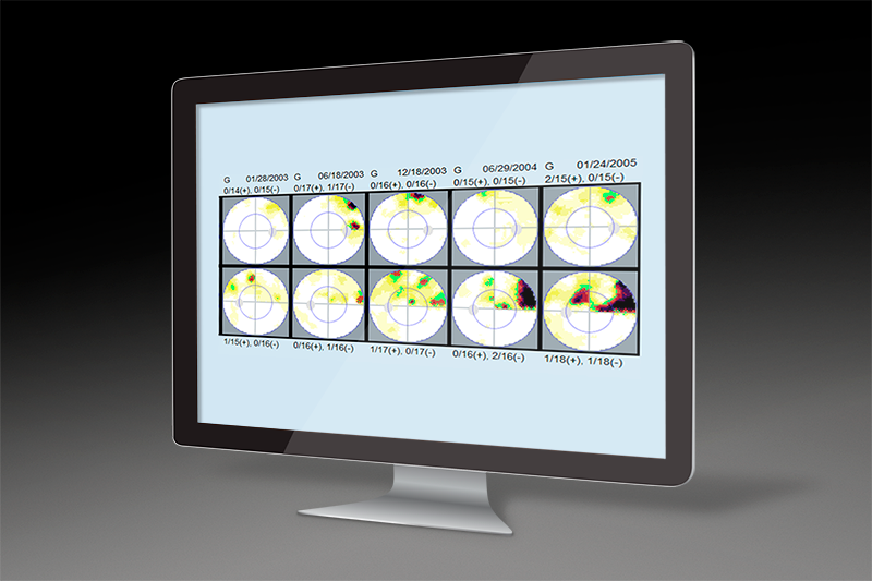
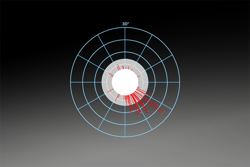
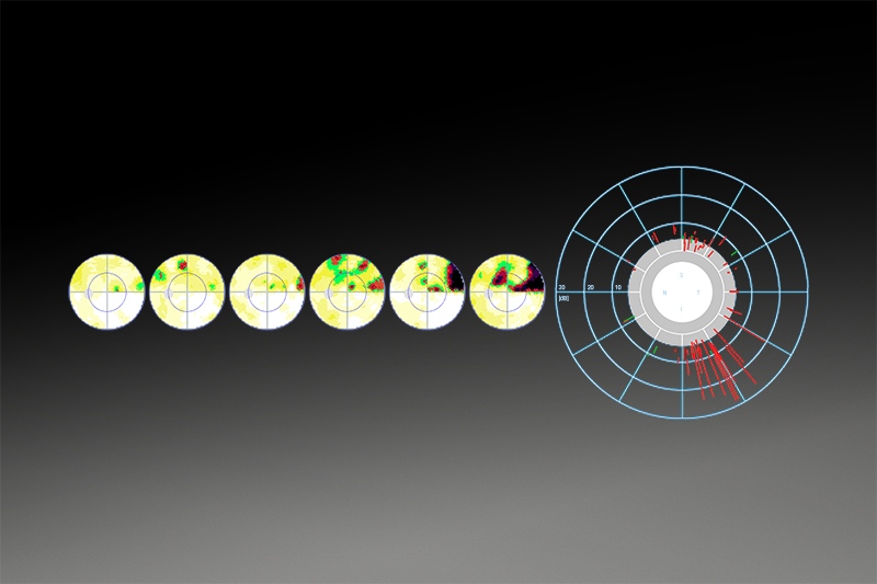
A clear view on glaucoma

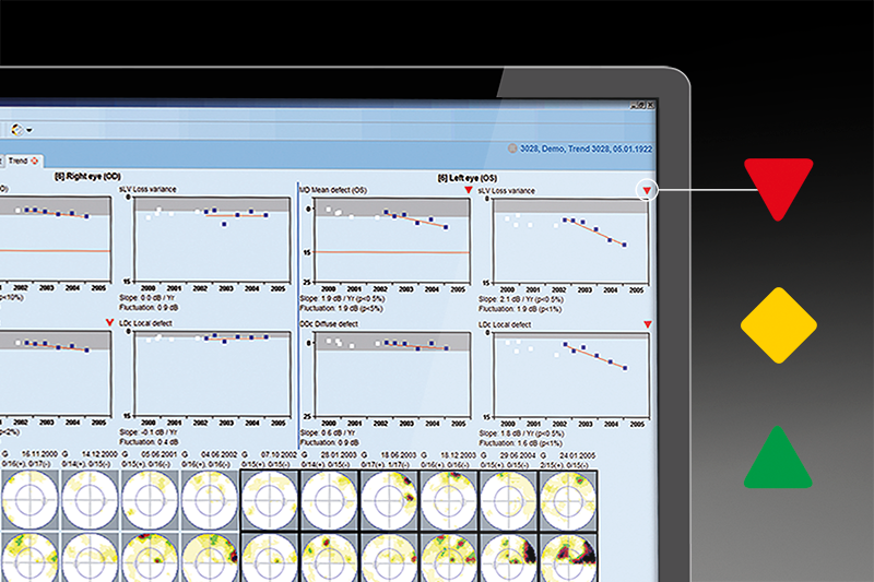
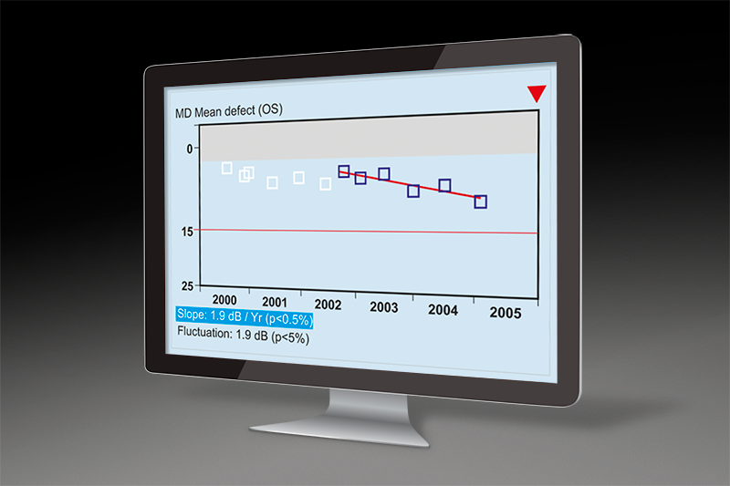
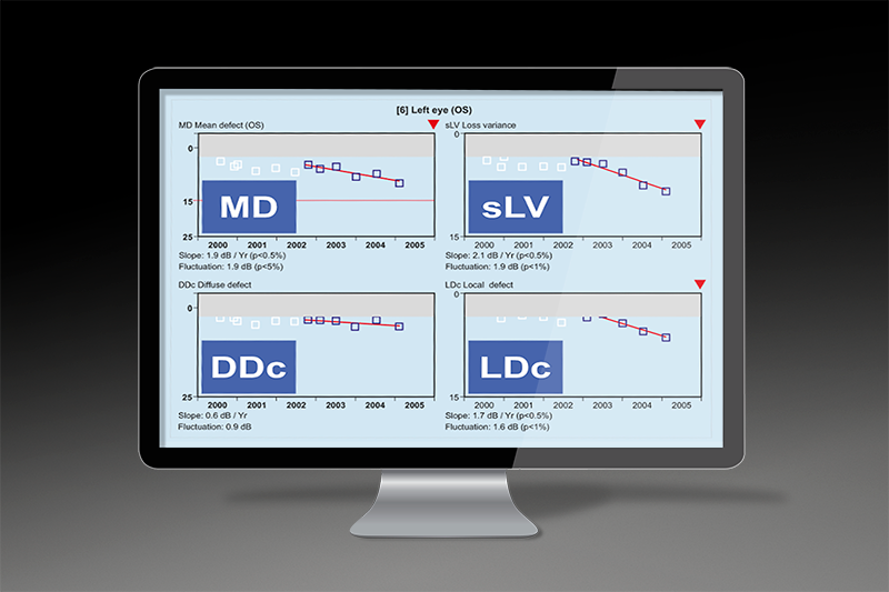
Get the most out of your glaucoma visual field with the highly sensitive Cluster Analysis, the intuitive Polar Analysis for structural comparisons and the easy-to-interpret EyeSuite Progression Analysis.

EYESUITE PROGRESSION ANALYSIS

Immediate identification of true change

Immediately identify levels of change with the EyeSuite Progression Analysis. It not only reveals whether change is significant, but also whether it is local or diffuse and how fast the change happening. For an effective clinical workflow, all results are displayed using intuitive graphical symbols and can be viewed directly in your office if the Octopus is networked.
EYESUITE PROGRESSION ANALYSIS

Assessing glaucomatous change from serial visual fields is not always easy, and expert agreement is moderate at best. Why not let EyeSuite Progression Analysis be your guide in assessing change? Red downward-pointing arrows show deterioration, green upward-pointing arrows show improvement and yellow diamonds show fluctuation at both at 1% and 5% significance level. This allows you to identify change at a glance.
The rate of glaucoma progression is key when choosing the right treatment for your patient. Therefore, EyeSuite Progression Analysis always shows you the rate of change in decibels per year. Now, you can quickly distinguish between fast and slowly progressing patients and adjust your treatment accordingly.

Looking at global visual field change is not always enough to fully assess visual field progression. Sometimes, diffuse loss due to cataracts overlaps with local glaucomatous change. In other cases, unspecific diffuse loss due to learning effects, fatigue or refractive errors, for example, can underlie small local changes. The indices Diffuse Defect DDc and Local Defect LDc are helpful in these cases. They quickly show you whether change is local, diffuse or both and further support your clinical decisions.
Are you looking for an effective way to encourage your patients' compliance? Then show them their visual field progression on the intuitive greyscale charts to help them to understand what glaucoma progression truly means for their vision. To do this effectively, network your Octopus perimeter and show the results directly on a computer screen in your examination room.
POLAR ANALYSIS/TREND

For intuitive structural comparison

Combining the results of both structure and function is key to obtaining a comprehensive assessment of the onset and progression of glaucoma. The Octopus Polar Analysis projects local visual field defects along the nerve fibers to the optic disk and displays them oriented as structural results. This makes structure-function correlation almost intuitive. Polar Analysis is available in both single field and trend view.

With Polar Analysis, the nerve fibre bundles that are in danger or defective are easily identified. Local defects are projected along the nerve fibres to the optic disk and are represented as red lines. The projected defects are vertically mirrored to look like a structural result and are scaled with rings for 10, 20 and 30 dB deviation.

Do you see glaucomatous progression in your structural results? Then consult Polar Trend to see whether you can find matching visual field deterioration. Polar Trend is based on the Polar Analysis. 
FIXATION CONTROL
FIXATION CONTROL

Perimetry you can trust

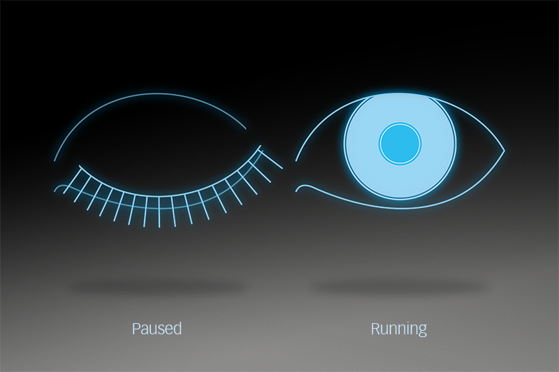
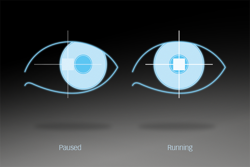
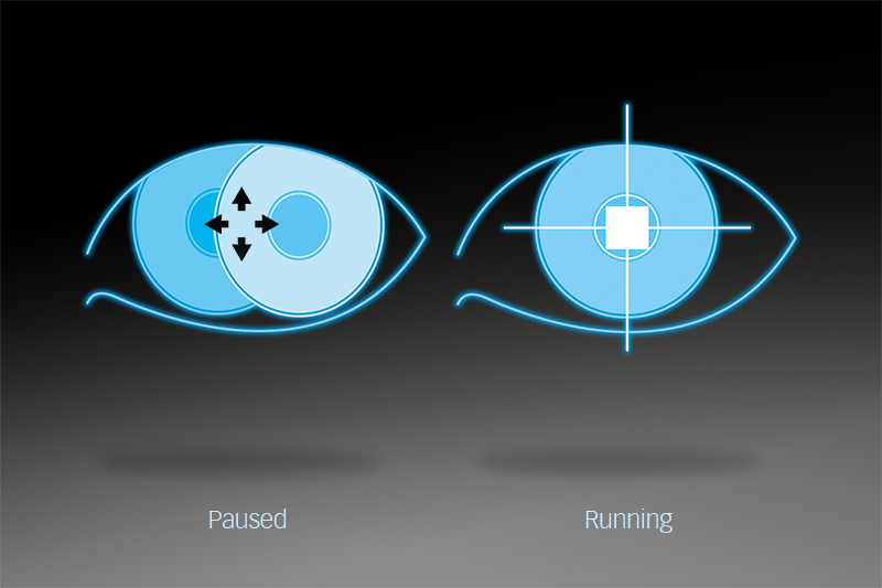
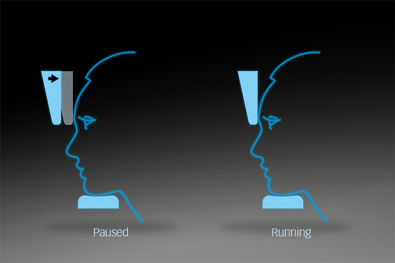
Enjoy the reliability of Octopus perimeters, for example with Octopus Fixation Control which automatically eliminates fixation losses from your visual field testing.

FIXATION CONTROL

Automatically eliminate fixation losses

Fixation losses due to low patient compliance are a major cause of unreliable visual fields. But you needn't worry any more. Fixation Control interrupts testing, when there is fixation loss, and automatically restarts, when the patient is properly fixating again. This ensures that each visual field point is reliably tested.
FIXATION CONTROL

Normal blinking prevents dry eyes and helps the patient to relax and concentrate during examination. With Octopus Blink Control, you need never worry again about missing a stimulus presented in static perimetry. Stimuli interrupted by the patient’s blinking are automatically repeated later during the test. This means that every test location is reliably tested.
Maintaining the correct pupil position during examination is essential for correct identification of the location of a defect. If the pupil position changes during stimulus presentation, due either to shifting of the head or eye movement, the Pupil Position Control automatically pauses the examination until the pupil is re-centred. The missed stimulus is automatically repeated later during the test. The result is a visual field that you can trust.

Remaining focused on the fixation target at all times is essential to prevent fixation losses. If patients rapidly move their eye to look for the next stimulus, Dart Control detects this fast movement and automatically interrupts the examination until a steady focus is achieved. Missed stimuli are automatically repeated later during the test making it easy to obtain reliable results.
Contact Control offers even more control and reliability by constantly assessing the correct patient position. If the patient pulls away from the instrument, it immediately notifies the operator and pauses the test to allow for re-positioning.
EYESUITE
EYESUITE

Easy integration in your practice workflow

The EyeSuite software has been designed for optimum patient flow in busy practices. It controls all Haag-Streit devices and allows for them to be networked with other Haag-Streit devices, your office computer and your EMR system without the need for any expensive third-party software.