HS HI-R 1000

A perfect combination

The HAAG-STREIT SURGICAL operating system HS 5-1000 consists of the SensoServo-driven HS Hi-R 1000 and the extremely steady floor stand FS 5-33. Enjoy highest optical quality with apochromatic optics delivering perfect color fidelity, strong contrast, and high resolution. The industry leading 25 mm stereo base provides superior 3D depth perception. A modular accessory structure fulfills all users’ wishes and the extraordinary stable and user-friendly carrying system allows easy positioning and intuitive operation with customized settings.

HS HI-R 1000
HS HI-R 1000

The masterpiece

“The optics on HS Hi-R 1000 are the best I have ever used. Additionally, it is extremely innovative as is apparent with the unique SensoServo Drive system which enable the user to move the scope with minimal effort, locks it in place immediately after moving, and avoids the need for balancing.”

SENSOSERVO DRIVE

Motorized Movement

The SensoServo system of HS Hi-R 1000 uses ‘fly-by-wire‘ technology to move the microscope in all directions with utmost precision and requiring minimal force. Though this technology also the balancing remains permanently stable even when changing the positioning of the microscope or e.g. shifting the stereoscopic observer scope from left to right.
SENSOSERVO DRIVE

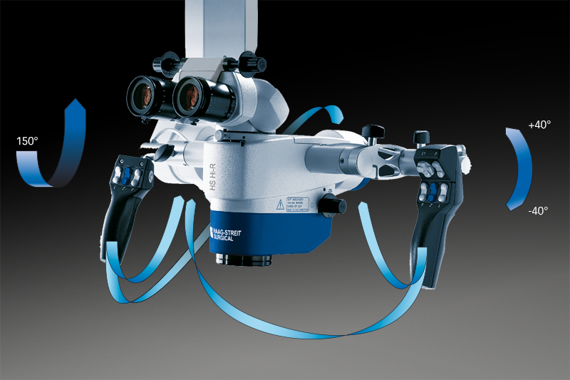
These characteristics are achieved via the SensoGrips that are activated as soon as the brake release buttons are pressed. They detect the pressure and control the motor movement in the microscope‘s joints accordingly. Individual movement characteristics can be chosen ranging from enhanced friction to feather-light.

FOCUSING

High flexibility

The variable focus assembly permits working distances from 224 mm to 510 mm without lens exchange. Optionally an alternative focus assembly is available that allows focusing at working distances of 200 mm to 450 mm. The integrated double iris diaphragm can be used to maximize the depth of field.
FOCUSING
 HANDLING
HANDLING

Full control at any time

In addition to the incorporated sensors, the hand switches allow control of focus, zoom, and light. Via a multifunction pad on the right SensoGrip, the microscope can be positioned without opening the brakes. Two programmable buttons can be individually set to control various functions.
FLUORESCENCE
FLUORESCENCE

Enhance visibility

Specific illumination and filtering can make the invisible visible when fluorescent effects are allocatable through relevant dyes. In the operating system HS 5-1000 HAAG-STREIT SURGICAL offers fluorescence equipment for ALA and ICG.

ALA/PPIX FLUORESCENCE

Intraoperative tumor visualization

ALA is used to visualize high-grade gliomas as well as the infiltrated areas adjacent to the tumor. The required filters for illumination (blue) and observation (yellow) integrated into HS Hi-R 1000 and are selected automatically when pressing the blue fluorescence mode button on the right hand grip. The surgeon can switch between the normal white light and the fluorescence mode at any time.
ALA/PPIX FLUORESCENCE

ALA, or 5-aminolevulinic acid, is a natural amino acid that the body metabolizes to heme, the red blood pigment. In high grade gliomas (grade III and IV), the last step of the heme synthesis is inhibited, which leads to an accumulation of protoporphyrin IX (PPIX) in the tumor cells. When illuminated with blue light, the PPIX emits a rich red fluorescence and marks the solid tumor with intensive red and the infiltrated areas with salmon-colored light.
ICG FLUORESCENCE

Intraoperative fluorescence angiography

ICG is used for intraoperative angiographies, particularly in neurosurgery for detecting stenoses, leakages, and aneurysms.
Following the injection of the ICG solution in the patient‘s bloodstream, the vessels become visible when the ICG flows by. Now, all irregularities can be seen on the unique M.DIS that is mounted in direct view of the surgeon and the HS MIOS displays.

ICG FLUORESCENCE
ICG FLUORESCENCE
ICG FLUORESCENCE

Equipped to function

Indocyanine green, abbreviated ICG, is a medical dye that emits infrared fluorescence when illuminated with light of the appropriate wavelengths. The microsurgical operating system equipped for ICG application includes a special infrared camera (in addition to the VIS camera) for detecting the ICG fluorescence and displaying it on the screen. Activation is easily controlled via the handles or the software.
ACCESSORIES
ACCESSORIES

Possibilities on demand

Depending on the demand, the operating microscope HS 5-1000 can be configured in a modular way. Various optional accessories are available.

MAGE INJECTION

Enrich your vision

Often, more information is needed than what hits the eye through the oculars of the operating microscope. In these situations extra information are provided in both oculars by our image injection system C.INJECT 1000. Featuring high definition, data can be injected in exceptional HD resolution into the oculars as an overlay to the live view or as a full image.
MAGE INJECTION
SECONDARY OBSERVATION

Flexibility

When working together the right accessory is needed:
  • Lateral observer scope with inclinable eyepiece head and image rotation for the assistant’s optimal comfort.
  • C.DUO offers face-to-face observation for two surgeons, lateral ports, and a separate camera connection. Eyepieces are fully rotatable for ergonomic positioning when tilting of the microscope.
  • To suit differences in height among surgeons various eyepiece heads allows best ergonomics.
HS MIOS 5

Comprehensive yet intuitive recording

The prime functions of HS MIOS 5 are the recording of operation scenes as well as the capturing and recording of snapshots, together with proper identification of patient and hospital data. Images and video streams can be stored on DVD-R/-RW, HDD, USB flash drive, and external USB hard disk drive or transmitted to the hospital PACS via DICOM.
HS MIOS 5
C.MOR HD / HD³
C.MOR HD / HD³

Compact HD camera

C.MOR HD is a full HD color video camera designed for the HAAG-STREIT SURGICAL microscope. Its ultra-compact camera head houses a 1/3” CMOS sensor that meets very high standards. In its 1 and 3-chip version, images are crisp and sharp. Different user settings allow individual adjustments.
M.FOCUS

Best focusing results

Automatic focusing is available with the HS MIOS 5 software module M.FOCUS. Benefit from best focusing results with an active focus area that is adjustable in size and position.


M.FOCUS
NAVIGATION
NAVIGATION

Open interface

The HS 5-1000 provides an open interface for connection with navigation systems. It supports pointer functions, image superimposition via C.INJECT 1000 as well as control of focus and positioning by the navigation system.

M.DIS AND C.MON HD

Display

Best control of the camera image and additional opportunities for observation are provided through monitors. Those may be mounted directly to the microscope head to serve the surgeon and the assistant  (M.DIS) or on the floor stand for further staff (C.MON HD).
M.DIS AND C.MON HD
MOUTH AND FOOT SWITCHES

Hands-free operation

The positioning of the microscope is normally operated using the handles. In addition, mouth and foot switches are also available. While the mouth switch allows the microscope to be moved during positioning, the smaller foot switch EF 2000 regulates focus and zoom settings. 14 partly programmable functions can be controlled with EF 5000 and the wireless EF 5001.

MAGNIFEYE

Seeing each detail

The MAGNIFEYE can be added to meet the highest demands in terms of up to 24x magnification. When using high magnification, you also benefit from the motorized X-Y movement HS Hi-R 1000 is offering for precise horizontal movements. These are easily controlled via the hand switch.

MAGNIFEYE
FS 5-33
FS 5-33

Solid as a rock!

The floor stand FS 5-33 integrates the latest technology with innovative damping for the lowest vibration. Even when fully equipped microscopes are mounted and the arm is stretched to its full length of 1870 mm, it still stays solid as a rock! Servo locks and state-of-the-art castors allow effortless maneuverability.

C.TAB AND CENTRAL TOWER

Control

The central tower of the stand neatly houses the light sources as well as accessories such as the recording system and camera control units. Control of the microscope system is accomplished with the C.TAB touch panel. It allows for setting the parking position, automated floor stand balancing, individual user settings for over 30 surgeons with 5 applications each.

C.TAB AND CENTRAL TOWER
IF GOLD DESIGN AWARD

Elegant and subtle with precise functionality

“Despite the products necessary complexity and high precision requirements, the design of this stand for surgical microscopes is very elegant and subtile. The device has a calm and clean appearance. This is a great advantage for daily use in hospitals as it helps to structure workflows in a more pleasant and stress-free manner. While the product is quite large it nonetheless features subtle forms and highly precise functionality. A truly outstanding and excellently designed surgical device.“

2X 300 W XENON

Powerful light

The two independent, powerful 300 W xenon light sources allow the best visibility, even in deep cavities. The quick exchange mechanism ensures the system is available quickly at all times. The illumination zoom HS Hi-R 1000 automatically adjusts the light to the size of the visible field and ensures optimal and constant brightness.

2X 300 W XENON
PARKING POSITION
PARKING POSITION

Compact for storage

The positioning for transport and parking is easily set and locked via C.TAB. The compact setting allows effortless maneuverability through hallways and requires little storage space.
CONNECTION PANEL

Easy accessibility

All needed connections for external monitors, network, or navigation system communication are easily accessible from one central panel.
CONNECTION PANEL