
The new paradigm in microsurgery
The advanced microsurgical operating system HS 3-1000 is based on the combination of the floor stand FS 3-43 with the operating microscope HS Hi-R 1000. It provides you with optimal working conditions as well as impressive modularity. The apochromatic optics and a unique stereo base of 25 mm ensure perfect color fidelity, strong contrast, and excellent depth perception. Electromagnetic brakes in the floor stand FS 3-43 support the smooth movements and minimize vibrations. Due to the stand’s long arm reach highest flexibility in the OR is achieved.

The masterpiece
“The optics on HS Hi-R 1000 are the best I have ever used. Additionally, it is extremely innovative as is apparent with the unique SensoServo Drive system which enable the user to move the scope with minimal effort, locks it in place immediately after moving, and avoids the need for balancing.”
Motorized Movement

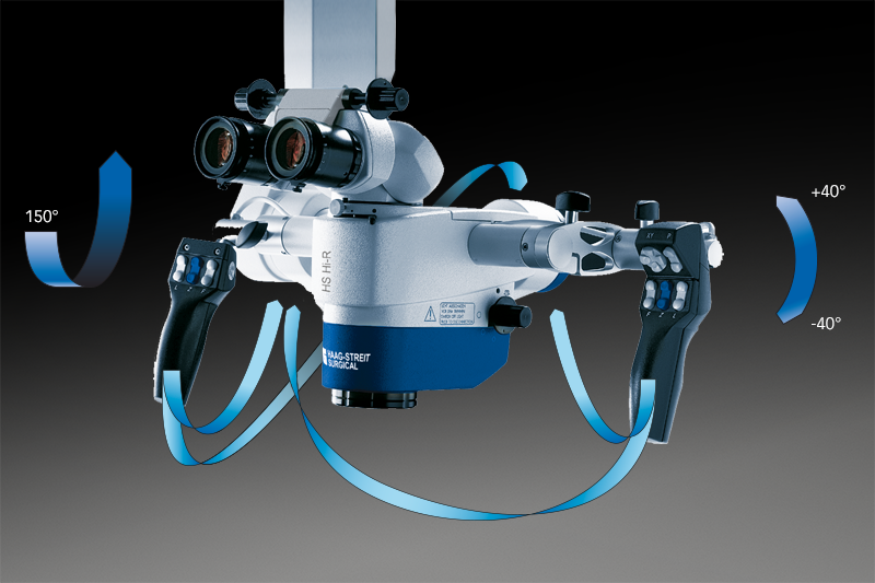
High flexibility


Full control at any time

Enhance visibility
Specific illumination and filtering can make the invisible visible when fluorescent effects are allocatable through relevant dyes. In the operating system HS 3-1000 HAAG-STREIT SURGICAL offers fluorescence equipment for ICG.
Intraoperative fluorescence angiography
Following the injection of the ICG solution in the patient‘s bloodstream, the vessels become visible when the ICG flows by. Now, all irregularities can be seen on the unique M.DIS that is mounted in direct view of the surgeon and the HS MIOS displays.


Equipped to function

Possibilities on demand
Depending on the demand, the operating microscope HS 3-1000 can be configured in a modular way. Various optional accessories are available.
Flexibility
- Lateral observer scope with inclinable eyepiece head and image rotation for the assistant’s optimal comfort.
- C.DUO offers face-to-face observation for two surgeons, lateral ports, and a separate camera connection. Eyepieces are fully rotatable for ergonomic positioning when tilting of the microscope.
- To suit differences in height among surgeons various eyepiece heads allows best ergonomics.


For improved treatment
Comprehensive yet intuitive recording


Compact HD camera
Best focusing results


Open interface
Enrich your vision


Display
Hands-free operation


Seeing each detail

Perfect reach, stability, flexibility
With its extreme arm length of 1600 mm the floor stand FS 3-43 allows great flexibility for positioning in the OR. The automatic balancing easily and fast adjusts the system when accessories are changed. Additional holders for foot switches, trays for camera control units, and a high resolution monitor or other accessories may be attached to the column.
At a glance

